Magnetic resonance imaging (MRI) is a diagnostic method capable of generating detailed three-dimensional anatomical images without exposing the patient to dangerous radiation. It is often used to detect and identify diseases, as well as to monitor ongoing treatments. It is based on a sophisticated technology that excites and detects the change of direction of the rotational axis of the protons present in the aqueous component of living tissues.
During an MRI exam, an electric current is circulated through coiled wires (coils) to create a temporary magnetic field on the patient’s body. Radio waves are sent and received by the device via a transmitter/receiver; these signals are used to create digital images of the scanned body area. An MRI scan typically takes 20 to 90 minutes, depending on the body part being studied.
For some MRI tests, drugs such as gadolinium contrast agents are given intravenously to change the contrast of the MRI image. These gadolinium-based drugs are based on rare metals; they are injected intravenously into the arm.
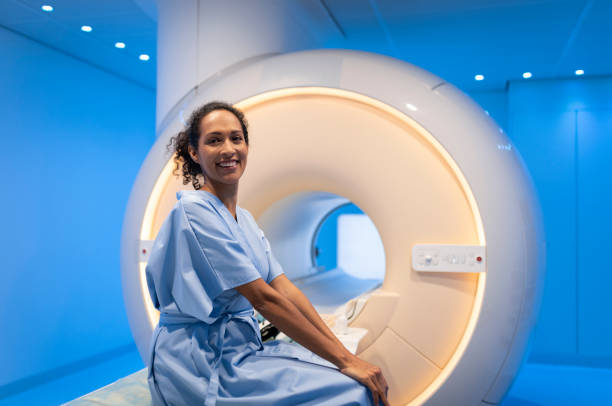
For the scan, the patient is made to lie down on a bed that slides inside the device, made in a tunnel. Scanning can take a long time, during which the subject must remain still. The examination is painless. The device is extremely noisy, so much so that the technician may suggest the use of earplugs.
WHAT IS SEEN WITH MRI?
In addition to these diagnostic uses, MRI can also be employed to guide some interventional procedures.
The exam is particularly suitable for the study of non-bony parts or soft tissues of the body. It gives information different from CT because it does not use the harmful ionizing radiation of X-rays. Brain, spinal cord, and nerves, as well as muscles, ligaments, and tendons, are much more evident on MRI than on radiological and CT scans, so this test is often required for knee and shoulder injuries.
In the brain, MRI can distinguish between white and gray matter and can also diagnose aneurysms and tumors. Since MRI does not use X-rays or other radiation, it is the imaging method of choice when frequent re-examination for diagnosis or therapy is required, especially for the brain. However, the method is more expensive than radiographic and CT techniques.
Functional magnetic resonance imaging (MRI) is a special type of MRI. It is used to observe brain structures and to determine which areas are activated (ie, consume more oxygen) during different cognitive activities. It is used to improve understanding of brain organization and offers a possible new standard for defining neurological status and neurosurgical risk.
RISKS
MRI images do not require the use of ionizing radiation, so patients are not exposed to the harmful effects of this energy source. However, although there are no known health risks associated with temporary exposure to the MR environment, the latter involves.
To have good quality images, the patient must remain very still for the duration of the procedure. Infants, young children, and others who are unable to remain calm must be sedated or anesthetized in order to perform the procedure. Sedation and anesthesia, in turn, have risks independent of the MRI procedure, such as slowing or difficulty in breathing and lowering blood pressure.
With contrast media, patients with severe renal insufficiency on dialysis are at risk of a rare but serious complication, nephrogenic systemic fibrosis, which appears to be linked to the use of some gadolinium-based means, such as gadodiamide.
Although a causal link has not been established, current U.S. guidelines recommend that gadolinium contrast agents be used in dialysis patients only when essential and that the examination is immediately followed by a dialysis session to promptly clear the contrast agent. from the body.
Although no effects on the fetus have been demonstrated, it is recommended that MRI be avoided as a precaution in pregnancy, especially in the first trimester when fetal organs are forming and contrast media, if used, could enter the fetal circulation.
Claustrophobia and panic attacks
Patients with even mild claustrophobia may have problems tolerating long scan times inside the device. Familiarizing yourself with the appliance and the process, as well as visualization, sedation, and anesthesia techniques, help overcome the discomfort.
Additional help can come from listening to music or watching a video or film, closing or covering your eyes, and having a panic button available.
Open MRI is an open-sided model, replacing the usually closed tube at one end; therefore it does not completely surround the patient. It was developed to meet the needs of people experiencing difficulties with the narrow tube and noises of the traditional version and those of patients whose body size makes the closed tube approach impractical. Open devices of the latest generation achieve high-quality images in many types of exams, but not all.
Patients with implants, external devices, and accessories
MRI presents specific dangers for patients with implants, external devices, and accessory medical devices.
An external device is an object that can touch the patient, such as an external insulin pump, brachial brace, or wound dressing.
An accessory device is a non-implanted medical device (a ventilator, a monitor, …), used to monitor or assist the patient.
The intense static magnetic field of the MR device attracts magnetic materials and can cause unwanted movement of the medical device.
Radiofrequency energy and magnetic fields that change over time can cause an implanted device and surrounding tissue to overheat, causing burns.
The magnetic fields and radiofrequency energy produced by the MRI device can also induce malfunctions of electrically active medical devices, interfering with the delivery of any therapies or functions.
The presence of the medical device degrades the quality of the image produced by the resonance, potentially reducing the information content and also leading to inaccurate diagnoses, with possible repercussions on the adequacy of medical treatment.
Patients with implanted devices, therefore, will not need to undergo MRI unless the device is specifically indicated as compatible (MRI-safe or MRI-conditional). An MRI-safe device is non-magnetic, contains no metals, does not conduct electricity, and does not cause harm in an MRI environment. An MRI-Conditional device can be used safely within an MRI environment that meets its specifications for safe use.
Any device with unknown MRI status should be considered MRI-incompatible.
Some inks used for tattoos contain metallic residues, although most are MR compatible. During the examination, notify the technician immediately if you experience discomfort or a sense of heat around the tattoo.
ADVERSE EVENTS
Adverse events following MRI are very rare, for example in the United States, where millions of tests are performed every year, the surveillance system receives about 300 annual reports of adverse events related to scans from manufacturers, distributors, laboratories, and patients resonance magnets. Most of these reports concern overheating and/or burns (thermal injuries). The most frequently reported problem is second-degree burns.
Other problems include bullet wounds (objects drawn to the appliance), fingers pinched or pinched by the patient couch, patient falls, and hearing loss or persistence of a sound ( tinnitus ) in one ear.
Sometimes, a jolting sensation from nerve stimulation may be felt due to the rapidly changing energy fields in the device.
USEFUL INFORMATION BEFORE UNDERGOING AN MRI
The space available during the MRI scan can be very cramped for patients sensitive to confined spaces, for obese people, or for particularly tall people. Inform the technician or doctor if you fear claustrophobic reactions.
During the scan, the device is extremely noisy and, to reduce the amount of noise, earplugs and/or protective headphones may be made available; in some cases, to make the examination less unpleasant, music earphones may also be worn.
If the MRI requires the injection of contrast medium, the technician inserts a small intravenous cannula into one arm. The injection of the medium can give a momentary sensation of cold. In any case, any pain or discomfort must be reported to the technician.
Remember that the examination was requested because the doctor believes that it can provide useful information for the management of one’s health. If you have any questions, don’t hesitate to ask them.
EXAM PREPARATION
On the day of the exam, the patient can drink, eat, and take medications as usual, unless otherwise instructed.
In some cases, it may be necessary to abstain from food and drink up to four hours before the exam, other times it is necessary to drink lots of water just before the MRI. It depends on the part of the body to be examined.
Upon arrival at the hospital, you are typically asked to fill out a form about your health and medical history. This data is needed by healthcare staff to ensure a safe examination. Be well informed on indications and contraindications to the examination.
Once you have completed the form, you will typically need to provide your written consent to the exam in order to proceed.
Depending on the body area to be examined, it may be necessary to wear a hospital dressing gown during the procedure. If not necessary, wear clothing without metal zips, buttons, hooks, belts, underwire (bra), clasps, or buckles.
Contrast medium
Some tests require the administration of the contrast medium. Contrast allows certain tissues and blood vessels to be seen more clearly and in greater detail.
In people with severe kidney problems, the contrast agent can cause damage to tissues and organs. If you have or have a history of kidney disease, a blood test may be done to understand kidney function and whether the procedure is safe.
It will also be essential to inform staff of any previous allergic reactions or clotting problems prior to injection of the medium.
Anesthesia and sedatives
MRI is a painless exam, so anesthesia (drugs that eliminate pain perception) is typically not required. In claustrophobic subjects a light sedative may be useful to relax; address the issue in time with your doctor before the exam.
In case of sedation, please note that it will not be possible to drive for the next 24 hours.
General anesthesia is often used in young children and infants; in these patients, it is necessary because it is essential that the patient remain perfectly still during the examination, which is often inexhaustible in young children when awake.